at Harvard Medical School
JET Q-FASTR Fund
October 2026
Since 2014, the Quadrangle Fund for Advancing and Seeding Translational Research (Q-FASTR) has accelerated early-stage research with potential for commercialization and, ultimately, the promise of improving people’s health. Since its inception, Q-FASTR has served as a critical nexus between basic science discoveries and innovative therapeutic possibilities. By providing funding, skills development, and project management, Q-FASTR removes obstacles to developing early-stage discoveries and creates new entry points for nascent technologies poised for commercialization. Q-FASTR buoys Harvard Medical School’s efforts to foster a research culture centered on therapeutic invention. Since the program’s launch, more than half of the laboratories on the Quad have sought Q-FASTR funding, indicating a growing and widespread interest in translation. By supporting HMS faculty with funding, resources, and partnerships, Q-FASTR has also laid the foundation for the Therapeutics Initiative, enabling researchers to demonstrate the merit and feasibility of their innovative ideas.

Q-FASTR by the Numbers
October X, 2026
Dear Bob,
I’m pleased to share an overview of the new Q-FASTR projects for fiscal year 2027, and to thank you once again for your crucial, foundational support of this transformative program. Q-FASTR has grown steadily in impact and reach over the years, becoming one of the world’s foremost pipelines from basic science research to commercialization and clinical impact, and an engine for innovation on the HMS Quad.
The projects selected for funding this year promise new treatments for specific illnesses such as type 1 diabetes and pulmonary arterial hypertension, as well as exciting new approaches to some of the most longstanding challenges our profession has sought to address, such as skin damage and pain itself. They represent the best medical science has to offer and exemplify our mission to improve health and well-being for all. In this report, each project is described by its principal investigator, offering a firsthand perspective on its promise and potential.
Thank you for your generosity, your vision in championing Q-FASTR, and your continued good counsel. I wish you a pleasant fall and hope to see you both soon.
Sincerely,

George Q. Daley, MD, PhD
Dean of the Faculty of Medicine
Caroline Shields Walker Professor of Medicine


George Q. Daley, MD, PhD
Extending CAR T Cell Therapy to Solid Tumors
Arlene Sharpe, MD, PhD
Kolokotrones University Professor, Harvard University
Chair, Department of Immunology, HMS
Vice Director, Gene Lay Institute
Eric Smith, MD, PhD
Director of Translational Research, Immune Effector Cell Therapies, Dana Farber Cancer Institute
CAR T cell therapy has transformed treatment for blood cancers but hasn’t yet worked well against solid tumors. Arlene Sharpe, MD, PhD, and Eric Smith, MD, PhD, are working to change that. They’re combining an engineered CAR T cell platform designed for solid tumors with a genetic screen that finds the internal brakes that wear these cells down.
The Challenge
Chimeric antigen receptor (CAR) T cell therapy, in which a patient’s own immune cells are re-engineered to recognize and destroy cancer, has proved genuinely transformative for blood cancers. Against solid tumors, it has largely failed: the engineered cells struggle to get into the tumor, and once there, they are quickly worn down. Sharpe’s project asks whether removing the cell’s internal “brakes” can close that gap.
The Approach
The project pairs two complementary strengths. The Smith lab has engineered second-generation CAR T cells against TROP2, a cell-surface protein widely expressed in solid tumors, including lung, breast, and bladder cancers. Those cells clear the small numbers of cancer cells left behind in non-small cell lung cancer (NSCLC) models, but they are only minimally effective against established, bulky tumors. The Sharpe lab has run CRISPR screens in living animals to find genes that act as natural brakes on the number and strength of T cells. Together, they are knocking out those regulators one at a time in TROP2-targeted CAR T cells and comparing the effect on expansion, killing capacity, and tumor control. The panel comprises CHIC2, PTPN2, RC3H1, TSC2, TNFAIP3, TGFBR2, and SOCS1, with TRAC as a negative control.
Progress to Date
The first milestone was technical and is now complete. The two labs merged their protocols, combining the Sharpe lab’s gene-editing step with the Smith lab’s method for delivering the CAR, given 2 hours apart. Critically, the two steps do not interfere with each other—editing does not reduce how well the CAR is delivered, and delivering the CAR does not reduce how well the edit works—making this a viable route to producing edited CAR T cells at scale. The team designed and validated guides against six genes of interest plus the TRAC control, reporting that the edits worked efficiently across the panel; validation of the SOCS1 guide awaits further work, as that stretch of DNA is hard to copy for testing.
With that platform in hand, the team ran two independent tests of how well the engineered cells killed tumor cells, against two human NSCLC lines, PC9 and HCC4006, using CD19-targeted cells, which do not recognize these tumors, as a control. In the first test, which counts surviving tumor cells at a set endpoint, no single knockout improved killing against both lines at both timepoints, but TSC2, TNFAIP3, and RC3H1 produced statistically significant improvements in at least one line at day 5. A second assay using live-cell imaging over 5 days showed few significant differences, because the unedited TROP2 CAR T cells were already killing almost everything, even at one CAR T cell for every two tumor cells. The team will test lower ratios of CAR T cells to tumor cells to leave more room to see a difference, and if that proves insufficient, will repeatedly expose the cells to their target to mimic the exhaustion CAR T cells experience during a prolonged fight against a tumor.
What Comes Next
The project is now moving into animal models, where the differences should become visible—culture assays cannot reproduce the physical barriers CAR T cells must overcome to infiltrate a solid tumor. Two studies are underway. The first is a pilot in the HCC4006 NSCLC model comparing five groups: no CAR T cell transfer, CD19 CAR T cells, non-targeting TROP2 CAR T cells, TSC2 knockout, and TNFAIP3 knockout, with tumor growth measured directly and body weight monitored for signs of toxicity. The second is a pooled competition study in which all eight knockout populations are transferred together into tumor-bearing hosts, then recovered from the spleen, the tumor, and the nearby lymph node, with each population counted by sequencing its edited gene. Because PTPN2, RC3H1, TGFBR2, and SOCS1 are already known to drive CAR T expansion in myeloma models, how well each population survives inside the tumor offers a direct way to rank them. Together, these should narrow the field to two or three candidates for tumor growth control studies and mechanistic analysis.

Engineered TROP2 CAR T cells rapidly eliminate lung cancer cells in a dish. Tumor cells grown alone (green) or with control CAR T cells that don’t recognize the tumor (blue) continue to grow. Tumor cells exposed to TROP2 CAR T cells, with or without additional gene edits (black), are almost completely cleared within about 40 hours. Because killing was this strong, the team will next test lower doses of CAR T cells to reveal which gene edits give the cells an extra edge.
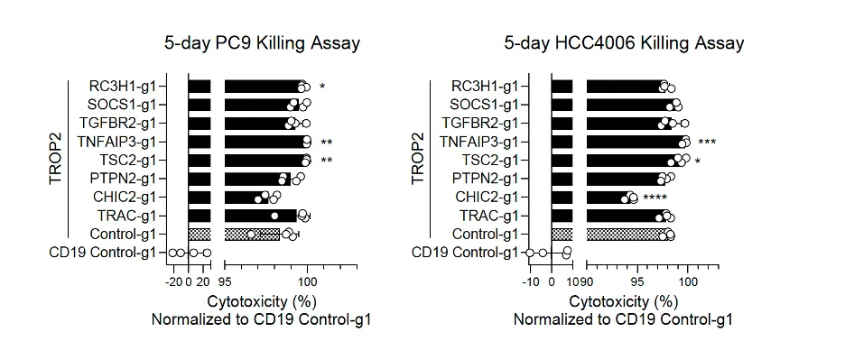
CAR T cells with specific genes switched off were tested for how well they killed two types of lung cancer cells over five days. Removing TNFAIP3 or TSC2 modestly improved killing (asterisks mark significant differences). These two genes will move forward into the team’s first study in mice.

Arlene Sharpe, MD, PhD

Eric Smith, MD, PhD
A Peptide and Its Receptor
—A New Lead for Alzheimer's Disease
Michael Greenberg. PhD
Nathan Marsh Pusey Professor of Neurobiology, HMS
Director of the Hock E. Tan and K. Lisa Yang Center for Autism Research, HMS
Professor of Neurology, Boston Children's Hospital
Josh Tycko, PhD
Postdoctoral Fellow, HMS
Michael Greenberg, PhD, and Josh Tycko, PhD, are following up on an unexpected hit from their high-throughput screening platform: a fragment of a protein linked to neurodegeneration that binds strongly to neurons. They have traced it to the receptor it acts on and are now exploring whether it could lead to a therapy for Alzheimer’s disease.
The Challenge
Secreted proteins and their cell-surface receptors account for roughly 70% of all Food and Drug Administration (FDA)-approved drug targets, yet the map of protein–cell interactions remains fragmentary, and there is no efficient way to predict which proteins will bind strongly to a given cell type. The Greenberg lab developed Barcoded Ligand Sequencing (BLS), a high-throughput platform that measures thousands of ligand–cell interactions in a single multiplexed experiment. Applied to primary neurons, BLS flagged a previously uncharacterized region of VGF—a protein fragment linked to neurodegeneration—as binding tightly to neurons. The project set out across three aims: pin down exactly which fragment is active, characterize its effects in mice, and identify the matching receptor on the cell surface.
Progress to Date
The team reports significant progress on the first aim and has largely completed the third. By systematically changing and deleting parts of the VGF peptides, they identified the three short stretches that drive the interaction with the receptor—work that also demonstrates that the platform can do this kind of fine-grained scanning directly on primary cells, which no existing method does well. They then used the strongest peptide as bait to isolate the long-sought VGF receptor, confirming it by lowering the receptor’s levels, raising them, and blocking it with a competitor across four cell systems, including primary neurons, with a separate technique confirming that the two bind in the test tube.
The receptor’s identity is what makes the finding compelling. It turns out to be a protein already independently implicated in neurodegeneration: it is the primary route by which neurons take up and pass along pathological Tau. Linking the VGF peptide to this receptor raises the possibility of a single unifying framework for understanding—and potentially intervening in—this disease pathway. The team validated the finding across four different cell systems, including primary neurons.
Reaching this result required a mid-course correction. The originally proposed CRISPR activation receptor screen worked as intended for a positive-control ligand–receptor pair but did not identify a receptor for VGF; the team pivoted to a mass spectrometry–based approach, which ultimately identified the receptor.
What Comes Next
A manuscript is near completion and will be submitted for publication and posted to bioRxiv. The team has updated its provisional patent on the BLS platform and will file a second covering the VGF peptides themselves. Remaining work on the first aim will characterize the downstream signaling and morphological consequences of VGF administration, and the second aim is beginning: testing the optimized peptide against disease progression in the 5xFAD mouse model of Alzheimer’s disease. The investigators note two constraints. Standing up the in vivo systems is slow, so they are running parallel in vitro models of Tau uptake and aggregation in the meantime. And the receptor’s signaling pathway is poorly characterized, appearing to depend more on the state of the neuron than other signaling molecules such as BDNF do, so they are testing VGF-mediated signaling across a wide array of cell treatment conditions. They are also applying to two Alzheimer’s-focused funding opportunities to support a more complete in vivo study, including Tau pathology, and to cross mice in which the receptor can be switched off with models of neurodegenerative disease, which would directly test whether this peptide–receptor interaction is necessary for the neuroprotective effect.
Michael Greenberg, PhD

Joshn Tycko, PhD


Using their barcoded screening platform, the team changed each building block of the VGF peptide, one at a time, and measured how well each version still bound to cells. Red squares mark changes that weakened binding, and blue squares mark changes that strengthened it. Clusters of red reveal the regions of the peptide that are essential for binding its receptor.
Small-Molecule Screens Against a New Tau Target in Alzheimer’s Disease and Related Tauopathies
John Flanagan, PhD
Professor of Cell Biology, HMS
Mark Namchuk, PhD
Puja and Samir Kaul Professor of the Practice of Biomedical Innovaton and Translation, HMS
Executive Director of Therapeutics Translation, HMS
Academic Director, Master of Medical Sciences in Therapeutic Sciences, HMS
Alzheimer’s disease and related tauopathies affect huge numbers of people, but there are very few promising drug targets. John Flanagan, PhD, and Mark Namchuk, PhD, are focusing on a newly identified regulator of tau hyperphosphorylation, a process that leads to the tau tangles that are a hallmark of these diseases, in search of new treatments.
The Challenge
Alzheimer’s disease is defined by two kinds of deposits in the brain: amyloid-β and an abnormally modified form of the protein tau. In 2025, the disease affected an estimated 7.2 million Americans at a cost of $384 billion. Immunotherapies against amyloid-β are approved, but their efficacy is limited, and there is good reason to think tau is the better target—yet no tau-targeting therapy exists. Flanagan’s laboratory has identified a novel regulator of tau hyperphosphorylation that sits at the top of the chain of events, raising the prospect that the entire cascade of tau pathology could be blocked by targeting a single protein. Proof of concept is already in hand: genetic and cell-biological experiments targeting the protein block neuron degeneration both in cultured cells and in mice.
The Approach
Working with Namchuk, the project is running two complementary searches for small molecules that change the regulator’s activity. The first is a DNA-encoded library (DEL) screen for compounds that bind the protein directly. The second is a FRET-based screen that tests compounds in living cells, using a light signal to detect changes in how the regulator interacts with its partner proteins. Follow-up tests measure the effect of candidate compounds further down the line, on tau itself. The overall goal is to establish the assays and identify compounds that can be carried forward into therapeutic development.
Progress to Date
The first stage of the DEL screen is complete. The advantage of the format is scale: a conventional chemical library holds on the order of 100,000 compounds, while a DEL screen can interrogate more than a billion, opening up a far larger region of chemical space. It also returns a different kind of answer. Rather than a handful of isolated hits, a DEL screen reports which chemical structures—and which combinations of them—bind the target. Based on those results, the team has selected 31 compounds to be remade in the lab to confirm they are chemically reliable before testing them in depth against the target proteins.
The FRET assay is being built in parallel. By fusing the target receptor to a fluorescent protein tag, the team is creating a proximity sensor that gives off a specific light signal when the receptor is engaged by its natural partners. That construct now expresses brightly and correctly at the surface of live human cells—the key first step for the assay.
What Comes Next
The 31 re-synthesized compounds will be tested in depth against the target proteins, with follow-up tests reading out the effects on tau itself. On the cell-based side, the team is optimizing the proximity sensor for automated, high-throughput screening, which will enable it to be used against conventional chemical libraries and, equally important, to validate the hits from the DEL screen.

Mark Namchuk, PhD
John Flanagan, PhD


Results from a screen of roughly 400 million compounds, each built from three interchangeable chemical parts (one per axis). Each dot marks a compound that bound the target. Compared with the negative control (far left), many compounds bound the target protein and a related protein. The team has selected 31 of these compounds to resynthesize and test further.

Live human cells in which the team’s target protein has been tagged with a fluorescent marker (yellow), confirming that it reaches the cell surface. This is the first step in building an automated, light-based assay to screen for drug candidates.
Office of Alumni Affairs and Development
25 Shattuck Street
Boston, MA 02115
